
Every shape a crystal could take – probing morphologies with Growth Explorer
Create and modify crystal shapes, and visualise growth elements on crystal faces
This blog introduces Growth Explorer, a new tool that helps scientists explore how crystal symmetry and intermolecular interactions influence crystal morphology and growth.
Why should you care?
We all love crystals. Right? And we all know how easy it is to grow them into the exact shape we want… Right? For those uninitiated in the dark arts of crystal growth, our beloved crystals like to test our patience by refusing to grow, transforming during or after growth, or appearing as a flurry of needles. We would all love to grow beautifully faceted prismatic blocky crystals. But alas, sometimes it’s just not meant to be. But it’s okay. It’s not you. It could just be the crystallographic constraints of your system. Let me explain.
Symmetry is at the heart of what is possible
Because of the symmetry relationships in the packing of a crystal, crystal morphology is an expression of that symmetry; in fact, before we had X-rays (before 1913), a common way to determine crystal symmetry was to measure interplanar angles – a potential exercise for the reader!
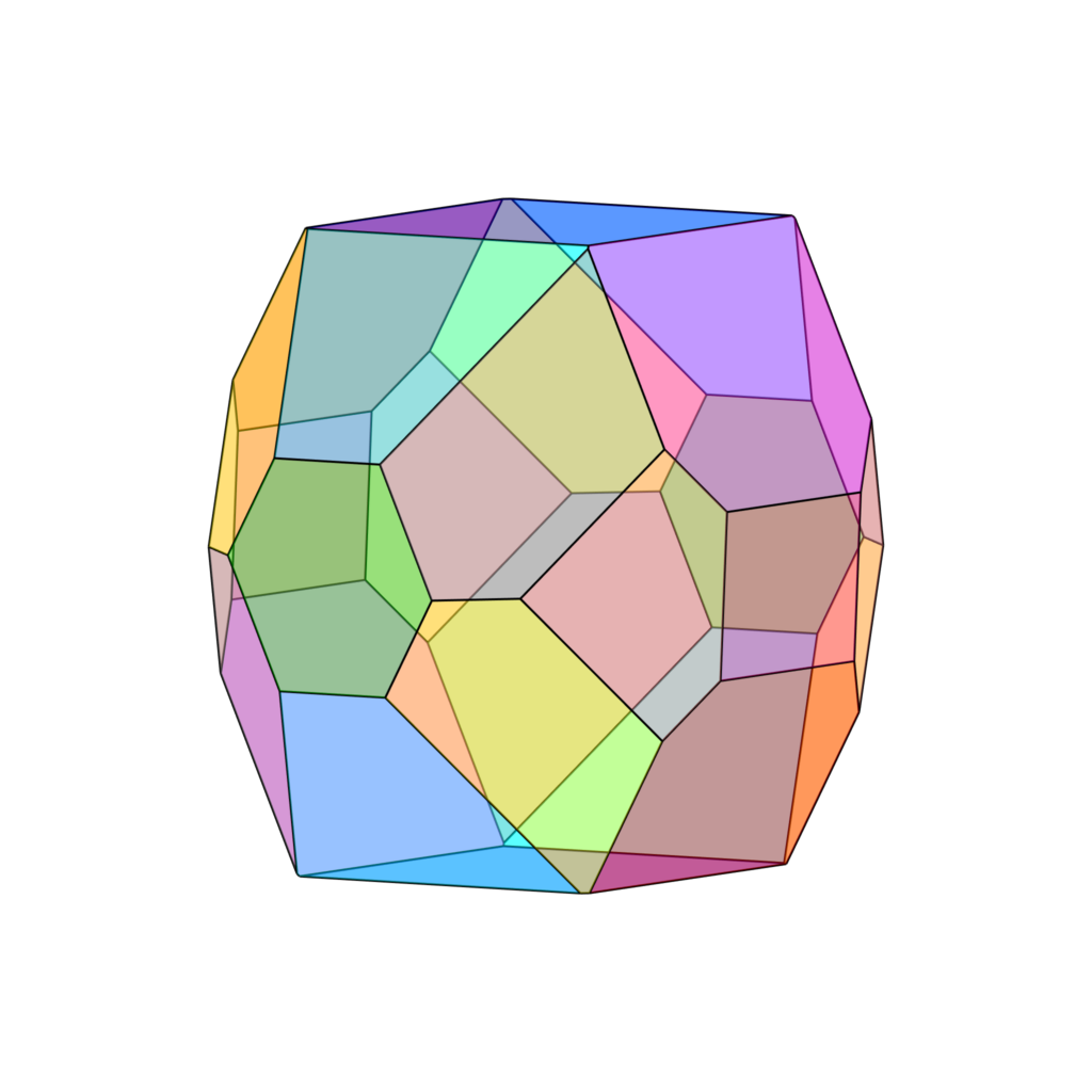
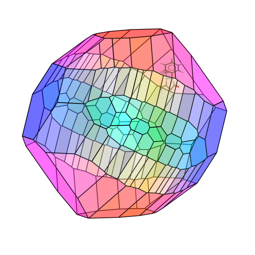
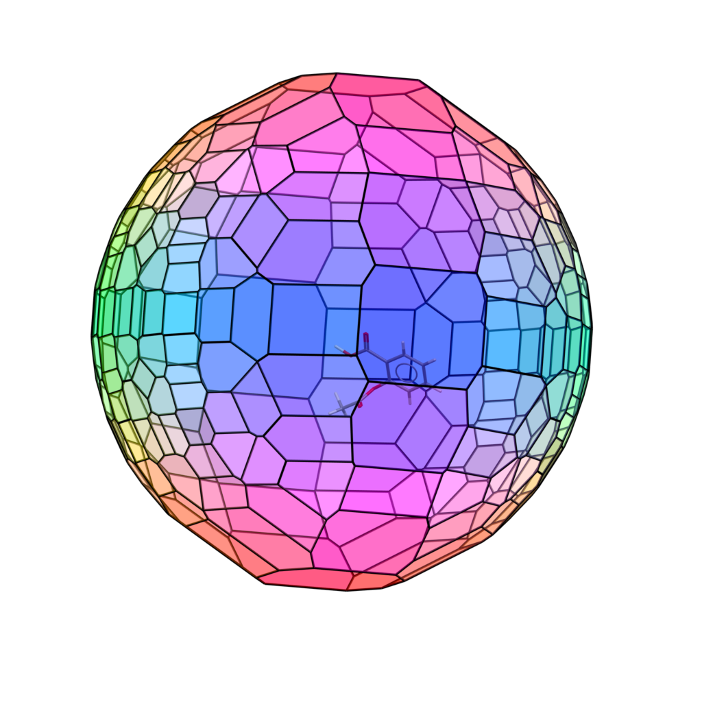
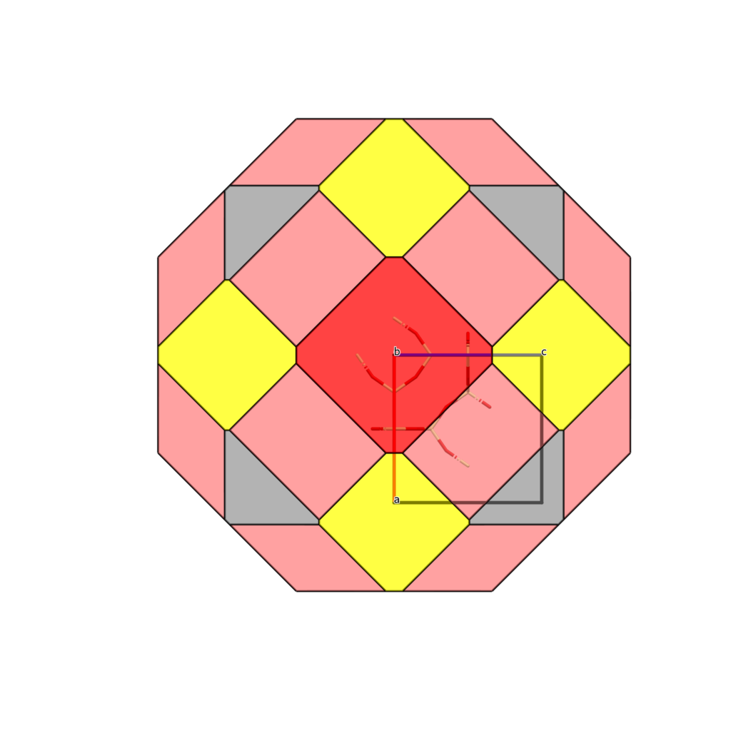
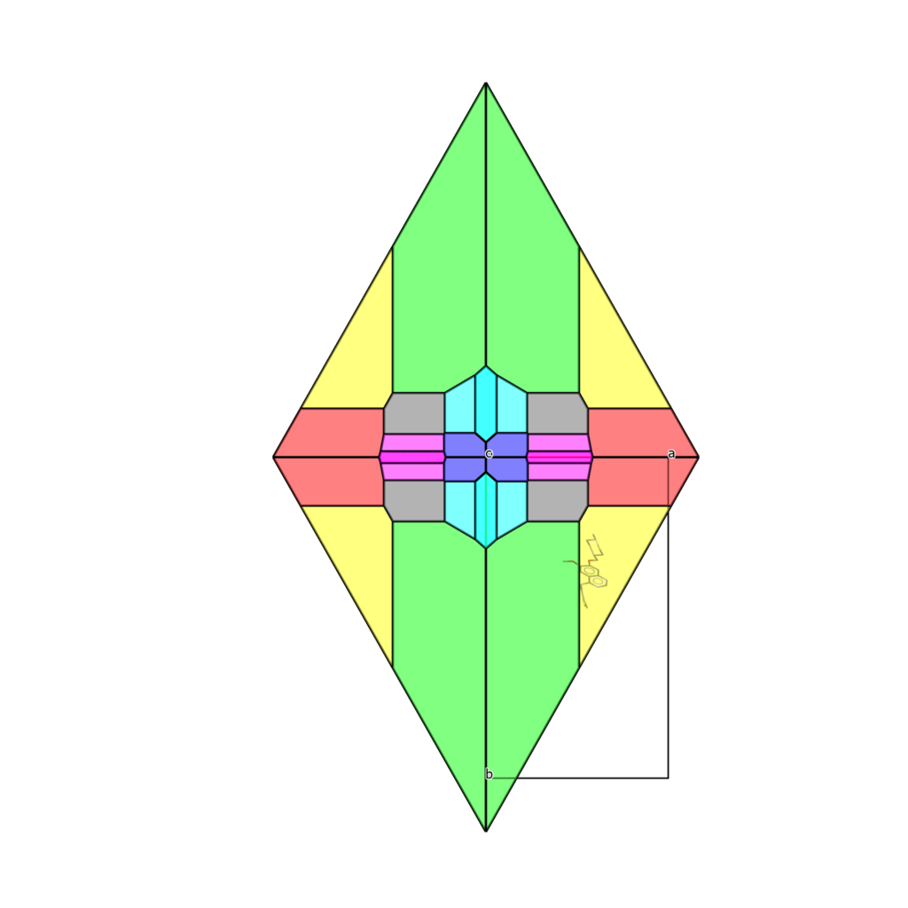
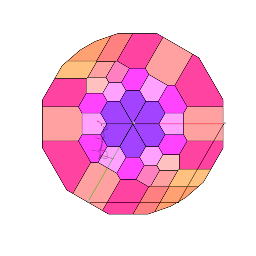
With Growth Explorer, you can now visualise all symmetry-allowed facets of a system. We colour the facets based on the {hkl} values(s) of each facet, mapping to the RGB (red, green, blue) colour model; i.e. (001) is a blue face where (100) is a red face. Below, we can see a couple of crystals where the space group shapes the available faces of the morphology; we call these Symmetry Allowed morphologies, and you can control how these are displayed by selecting either the maximum hkl order or the minimum inter-planar distance (d-spacing).
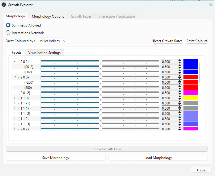
The Growth Explorer dialog allows you to see the symmetry relationships between facets and modify the relative growth rate for each facet or form so you can explore different shapes.
Can you guess the space groups of these symmetry-allowed morphologies?
But energetics also matter
Since I am sure you are an avid reader of our blogs, you will have seen our post on the addition of Periodic Bond Chains through Interaction Network Analysis. We’ve plugged those interactions into this tool, so you can now explore the faces defined by the energy holding the crystal together. The face selection is based on the Periodic Bond Chains (PBCs) of classical Hartman-Perdok theory [1] and shows which faces those interactions could plausibly support. The tool breaks each face down into growth layers, step edges, and individual kink sites where molecules attach, each with its own interaction energy. This does not predict the final morphology you will see in your crystallisation experiment, but it will narrow down the potential faces observed/shown, and you can explore how a particular pattern of molecular interactions could translate into a particular shape and to test “what ifs”.
What does a flat face look like up close? Here is a layer on the (001) face of NAPHTA52. The grey molecules are the layer beneath it; the coloured connections show intermolecular interactions, with colour indicating their strength. From above, you can trace the network across the face. Turn it on its side and you can see the new layer sitting on the crystal below. The PBCs describe connected directions within the structure; where at least two run in the plane of a face, they can support a flat growth layer.
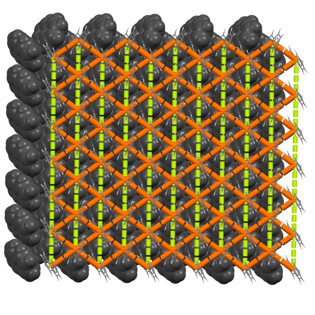
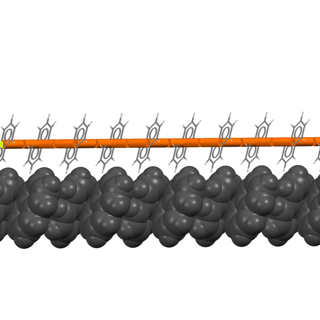
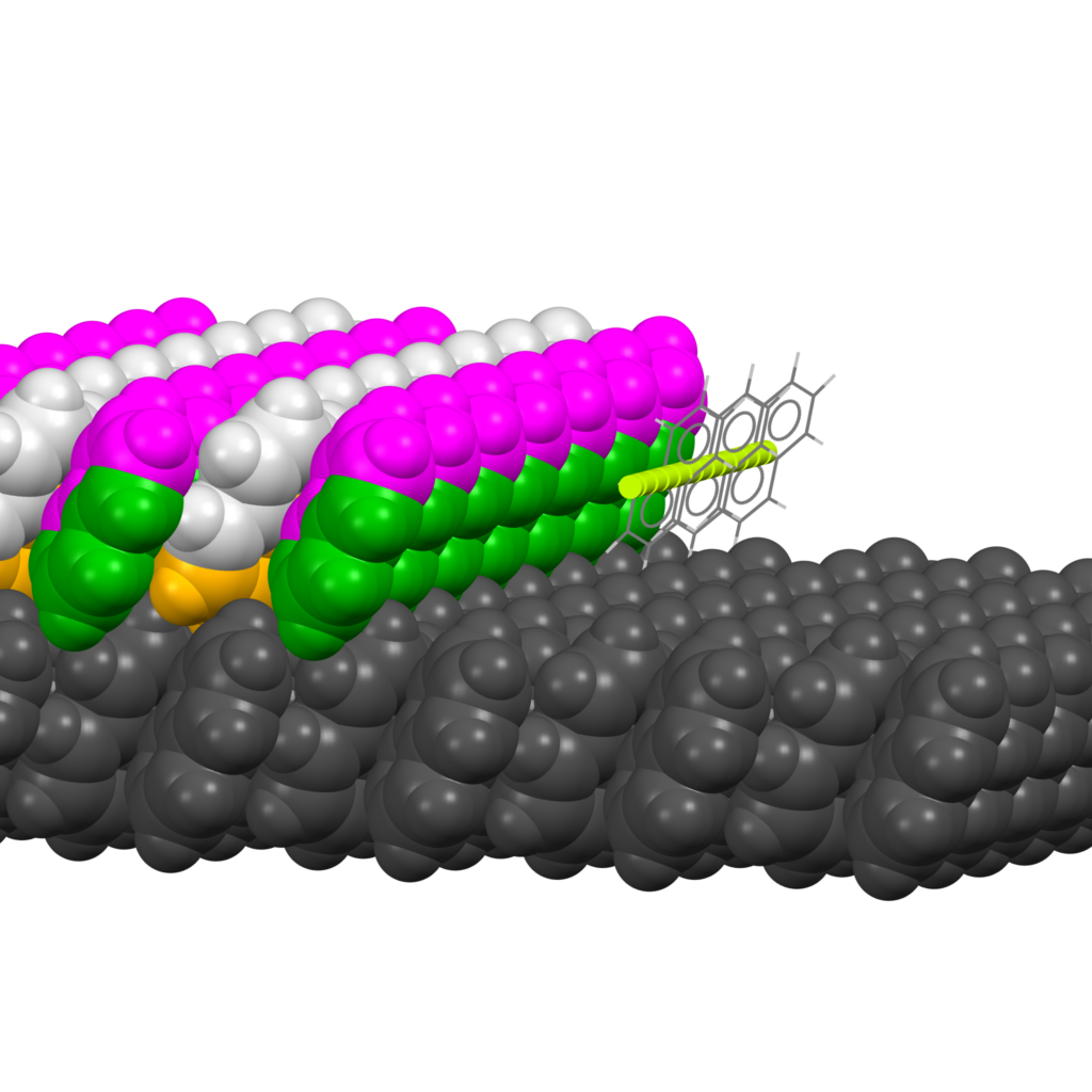
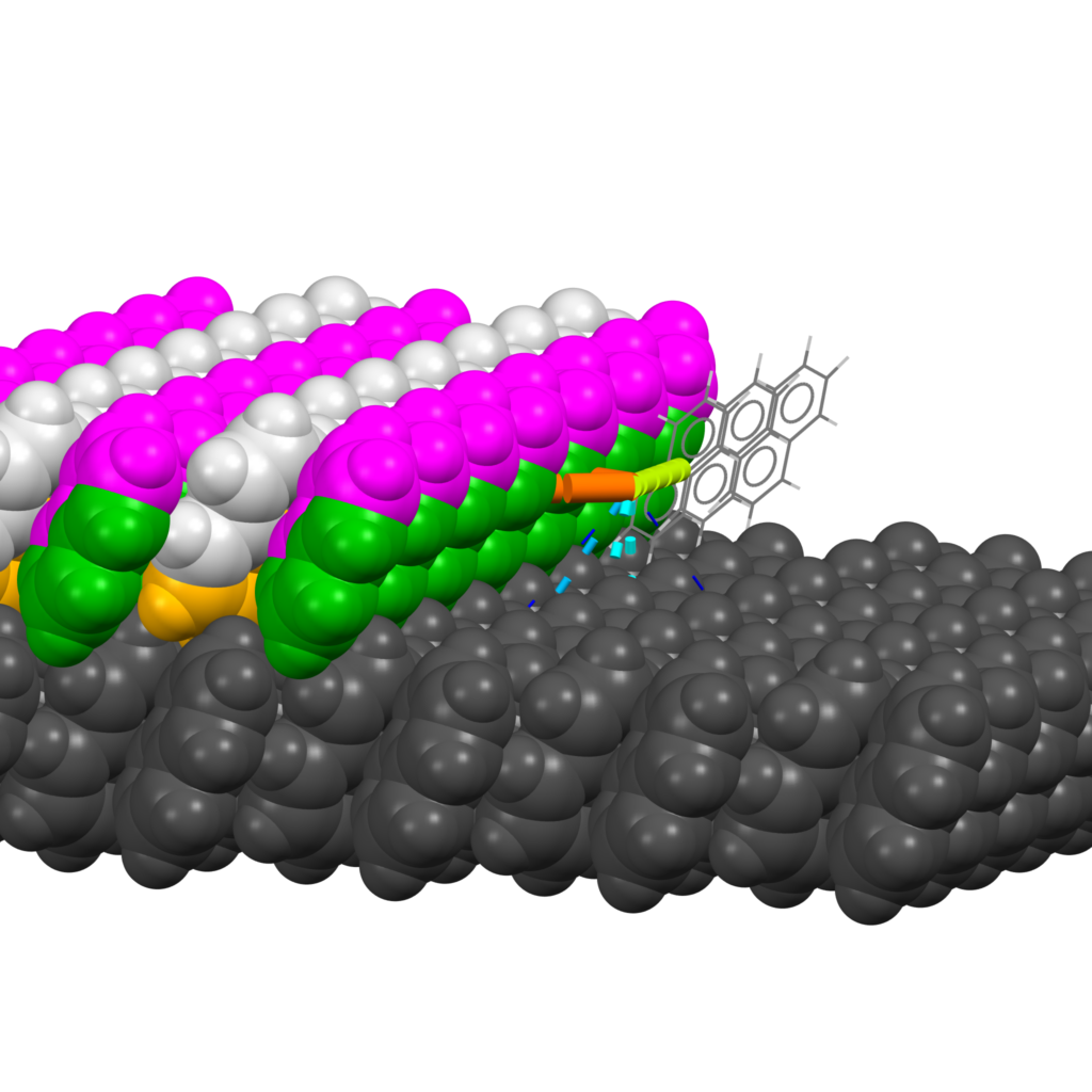
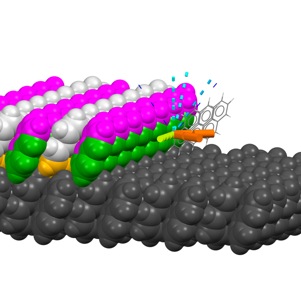
A layer does not have to appear all at once. Imagine it spreading across the face; its unfinished boundary is a step edge, built from rows of molecules. A new molecule can join at a kink site at the end of one of those rows. The energies reported for each layer, step, or kink are sums of the intermolecular interactions broken to create it.
The three views below pick apart the interactions around a kink site: along the row, back into the bulk crystal, and out towards the surrounding environment. Comparing these contributions helps you see what holds a growth unit in place, and why changing the interactions might change how readily a layer grows. Think of it as a closer look at the possibilities, as opposed to predicting the crystal growth.
The Growth Explorer functionality is available in Mercury and the CSD Python API to users with a CSD-Particle or academic CSD-Enterprise licence.

The Growth Explorer functionality was developed as part of the DigiCCAMMS program, in partnership with the University of Durham. This project will integrate the ADDICT prototype [2] into the CSD-Particle suite using Innovate UK funding as part of the Sustainable Medicines Manufacturing Innovation: Collaborative R&D competition.
References
[1] (a) Hartman, P.; Perdok, W. G., Acta Cryst 1955, 8 (1), 49–52. On the relations between structure and morphology of crystals. I (b) Hartman, P.; Perdok, W. G., Acta Cryst 1955, 8 (9), 521–524. On the relations between structure and morphology of crystals. II (c) Hartman, P.; Perdok, W. G., Acta Cryst 1955, 8 (9), 525–529. On the relations between structure and morphology of crystals. III
[2] Li, J.; Tilbury, C. J.; Kim, S. H.; Doherty, M. F., Progress in Materials Science 2016, 82, 1–38. https://doi.org/10.1016/j.pmatsci.2016.03.003